 Improved transformation efficiency of group A Streptococcus by inactivation of a type I restriction modification system
PLoS ONE


Competing Interests: The authors have declared that no competing interests exist.

- Altmetric
Streptococcus pyogenes or group A Streptococcus (GAS) is a leading cause of bacterial pharyngitis, skin and soft tissue infections, life-threatening invasive infections, and the post-infectious autoimmune syndromes of acute rheumatic fever and post-streptococcal glomerulonephritis. Genetic manipulation of this important pathogen is complicated by resistance of the organism to genetic transformation. Very low transformation efficiency is attributed to recognition and degradation of introduced foreign DNA by a type I restriction-modification system encoded by the hsdRSM locus. DNA sequence analysis of this locus in ten GAS strains that had been previously transformed with an unrelated plasmid revealed that six of the ten harbored a spontaneous mutation in hsdR, S, or M. The mutations were all different, and at least five of the six were predicted to result in loss of function of the respective hsd gene product. The unexpected occurrence of such mutations in previously transformed isolates suggested that the process of transformation selects for spontaneous inactivating mutations in the Hsd system. We investigated the possibility of exploiting the increased transformability of hsd mutants by constructing a deletion mutation in hsdM in GAS strain 854, a clinical isolate representative of the globally dominant M1T1 clonal group. Mutant strain 854ΔhsdM exhibited a 5-fold increase in electrotransformation efficiency compared to the wild type parent strain and no obvious change in growth or off-target gene expression. We conclude that genetic transformation of GAS selects for spontaneous mutants in the hsdRSM restriction modification system. We propose that use of a defined hsdM mutant as a parent strain for genetic manipulation of GAS will enhance transformation efficiency and reduce the likelihood of selecting spontaneous hsd mutants with uncharacterized genotypes.
Introduction
As part of our research into molecular pathogenesis of infections due to group A Streptococcus (S. pyogenes or GAS), our laboratory routinely constructs mutant strains of GAS using allelic exchange mutagenesis by transformation of a wild type strain with a temperature-sensitive plasmid carrying the desired mutation. During characterization of one such mutant strain, we made the incidental discovery that the mutant harbored a 694 bp deletion in hsdM, which encodes a component of a type I restriction modification system. The deletion resulted in a frameshift mutation at codon 123, altering the remainder of the protein sequence and introducing a premature stop codon at amino acid 170. As discussed below, because inactivation of this system can be associated with enhanced susceptibility to transformation with foreign DNA, three questions arose concerning inactivating mutations in hsdM or in genes encoding other components of the restriction modification system: (1) Does inactivation of hsdM result in increased transformation efficiency? (2) Does the process of transformation select for spontaneous inactivating mutations in the restriction modification system? (3) Could deliberate disruption of the system be exploited to facilitate introduction of foreign DNA for genetic manipulation of GAS? The current investigation was undertaken to answer these questions.
Type I restriction modification systems are comprised of three subunits that work together to recognize and cleave intracellular foreign DNA at specific sites and also to protect host cell DNA from cleavage by methylating it at the same recognition sites. Together, the three protein subunits comprise the “host specificity of DNA” system, or Hsd. The three enzymes of the Hsd system include the specificity subunit, HsdS, the restriction subunit, HsdR, and the modification subunit, HsdM. HsdS requires interaction with HsdM in order to bind to DNA. Once bound, the HsdS/HsdM complex can interact with HsdR. HsdR can then exert endonuclease activity at specific recognition sites, activity which is dependent on the methylation state of the target sequence. These functions of the three enzymes are tightly linked, as interaction with HsdM is necessary both for HsdS to bind to DNA and for HsdR endonuclease activity to occur [1]. It has been postulated that HsdM serves as a probe to detect methylated adenine residues on the target DNA. Only when methylation is absent on both strands can HsdM enter the major groove and confer the required conformation for HsdR to function. Together, the three-protein complex is able to cleave foreign DNA after it enters the cell as a type of bacterial defense system.
The Hsd system in GAS is a typical type I restriction modification system. It efficiently targets and cleaves foreign DNA that may enter the cell either naturally or artificially through electroporation. Accordingly, GAS is notoriously difficult to manipulate genetically in the laboratory setting. Previous studies have reported that inactivation of the Hsd system in GAS is associated with increased transformation efficiency [2,3]. One study done in an M28 strain found that deletion of the three components of the Hsd system was associated with increased transformation efficiency [2]. Another study characterized a group of emm1 clinical isolates associated with invasive infections. These strains were found to harbor a spontaneous deletion that included part of hsdR, the entire hsdS and hsdM gene sequences, and a portion of an adjacent two-component system. Strains with the deletion were shown to have increased transformation efficiency compared to contemporaneous emm1 isolates without the deletion [3].
In this study, we introduced a large deletion in hsdM in a GAS strain representative of the M1T1 clonal group widely implicated in invasive GAS disease. We found that inactivation of hsdM resulted in increased transformation efficiency of the strain, and that the mutation was not associated with a significant change in transcript abundance for other GAS genes including those encoding key virulence factors. In addition, we observed that the process of transformation selects for strains that have acquired spontaneous mutations in the Hsd system. We suggest that using a defined hsdM mutant as the parental strain for genetic manipulations could improve transformation efficiency and reduce the likelihood that transformation would select for an undefined spontaneous mutation in the restriction modification system with unknown potential off-target effects.
Results
Inactivation of hsdM results in increased transformation efficiency
Since restriction modification systems target foreign DNA for destruction by the host cell, we hypothesized that inactivation of the Hsd system by deletion of hsdM would result in a strain with increased transformation efficiency. To test this hypothesis, we introduced a 1,005 bp in-frame deletion into the hsdM coding sequence of GAS strain 854, an emm1 isolate representative of the globally prevalent M1T1 clonal group that is dominant among GAS emm1 strains associated with invasive infections [4]. The mutation of hsdM was confirmed by DNA sequencing. No growth deficiency was observed in a growth curve compared to wild type strain 854 grown in THY medium. We tested transformation efficiency by electroporation of wild type strain 854 or the hsdM knockout strain 854ΔhsdM using 1 μg of plasmid DNA. We then serially diluted the transformation reactions, plated the dilutions on selective agar, and counted the number of transformants that resulted from each reaction. These experiments revealed an increase in transformation efficiency of the ΔhsdM strain of approximately 5-fold compared to the wild type strain (Fig 1).

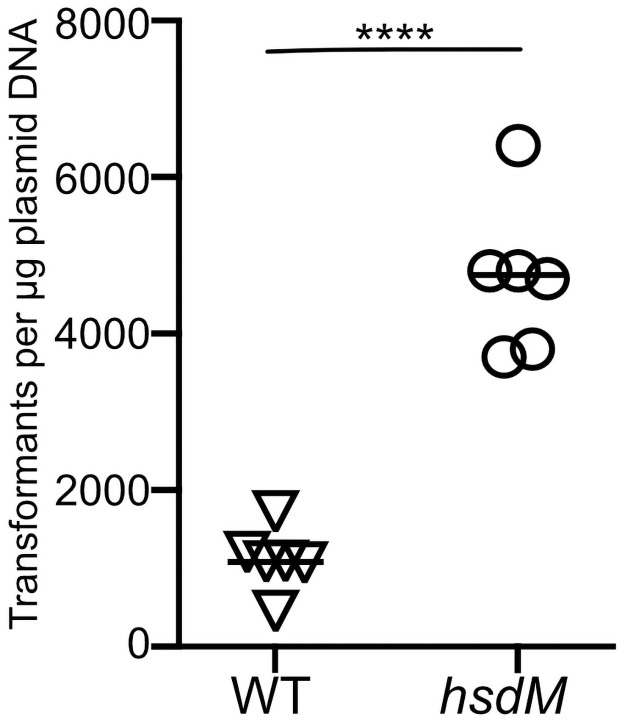
Deletion of hsdM results in increased transformation efficiency.
Cells of wild type GAS strain 854 (triangles) or mutant strain 854ΔhsdM (circles) were subjected to electroporation with 1 μg of plasmid DNA and then serially diluted and spread on selective medium for quantitative culture. Data points represent independent experiments; horizontal bars indicate the mean. ****P<0.0001, Student’s t-test.
Transformation of wild type GAS selects for mutants in hsdRSM
Because inactivation of HsdM resulted in a higher transformation rate, we wondered whether the process of transformation of wild type GAS might select for bacterial cells that had acquired a spontaneous inactivating mutation in the Hsd system. We screened for such mutations by sequence analysis of the hsdM locus of ten independently derived mutant strains of GAS 854 that had been previously transformed during allelic exchange mutagenesis of various genes. We found that four of the ten had acquired spontaneous mutations in the hsdM gene. These mutations included a deletion encompassing the entire hsdM coding sequence, a 1.5kb insertion, a missense point mutation, and a nonsense mutation (Table 1). Sequence analysis of the entire hsdRSM locus revealed that two other strains among these ten had mutations in either hsdR or hsdS (Table 1). Thus, including the mutations in hsdM, we found spontaneous mutations in the hsdRSM locus in six of the ten strains that had been previously transformed. Five of the six mutations introduced premature stop codons and/or large insertions or deletions in coding regions, strongly suggesting that these mutations all result in inactivation of at least one component of the Hsd system. By modeling the structure of HsdM to the published structure of a homolog in E. coli (PDB accession number 7BST) [5], we mapped amino acid T316 to the interface between HsdM and HsdR (Fig 2). The corresponding residue in the E. coli HsdM, N319, has been proposed to interact with R456 of HsdR, which aligns with K449 in the GAS HsdR sequence. Thus, mutation of GAS T316 from threonine to proline in the CsrS D148N E151Q D152N strain may disrupt the interaction of HsdM with HsdR and impact the function of the enzyme complex.
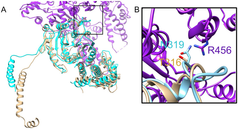
Modeling of GAS HsdM.
(A) Modeled structure of HsdM from GAS is shown in brown superimposed on a portion of the published structure of HsdR2M2S1-OcrIS from EcoR124I (PDB 7BST). E. coli HsdM is shown in teal, and HsdR is shown in purple. (B) Close up view of boxed section of panel A showing the interface between HsdM and HsdR. Proteins are in ribbon representation with residues R456 from HsdR, N319 from HsdM (EcoR124I) and the corresponding residue T316 from HsdM (GAS) shown in stick representation. Alignment has a normalized Z-score of 3.30.

| Strain | hsd mutation |
|---|---|
| 854ΔcsrR | 694 bp deletion in the coding region of hsdM |
| 854CsrS H280A | Premature stop codon at position 380 (of 526) in hsdM |
| 854Slo Y255A | 1.5 kb insertion in the coding region of hsdM |
| 854CsrS D148N E151Q D152N | T316P substitution in HsdM |
| 854CsrS T284A | 4 nucleotide insertion duplication that introduces a premature stop codon at position 794 (of 992) in hsdR |
| 854NADase G330D | 621 bp deletion in the coding region of hsdS resulting in a deletion from E160 to G367 |
| 854Δslo | none |
| 854Δnga | none |
| 854ΔcsrS | none |
| 854speB::Ω | none |
Since each of the spontaneous mutations we observed in transformants of strain 854 is different, we infer that the various mutations in the hsdRSM locus arose as independent events, rather than a single ancestral mutation in the parental wild type strain. Taken together, these observations strongly suggest that GAS isolates that have been successfully transformed in the laboratory setting may have a mutation that inactivates the Hsd system, and that the process of transformation selects for such mutants.
Deletion of hsdM in emm1 GAS strain 854 is not associated with altered expression of virulence genes
To assess the impact of inactivating hsdM in strain 854, we performed RNA-seq to evaluate global gene expression changes in the ΔhsdM strain compared to wild type. Twelve genes exhibited a 2-fold or greater change in transcript abundance for the mutant compared to wild type (Table 2). Assessment of gene expression by qRT-PCR of these twelve genes failed to confirm a significant change in expression of any of the genes (Table 2).
| Gene Locus | Gene Name | Gene product | RNA-seq | qRT-PCR |
|---|---|---|---|---|
| M5005_Spy_1066 | amyB | neopullulanase/cyclomaltodextrinase/maltogenic alpha-amylase | 1.277 | -0.327 |
| M5005_Spy_1067 | malX | maltose/maltodextrin-binding protein | 1.197 | 0.061 |
| M5005_Spy_1399 | - | PTS system galactose-specific transporter subunit IIC | 1.179 | -0.576 |
| M5005_Spy_1499 | grpE | putative Hsp-70 cofactor | 1.421 | 0.160 |
| M5005_Spy_1634 | lacF | PTS system lactose-specific transporter subunit IIA | 2.048 | -0.066 |
| M5005_Spy_1635 | lacD.2 | putative tagatose 1,6-diphosphate aldolase | 1.657 | -0.004 |
| M5005_Spy_1636 | lacC.2 | putative galactose-6-phosphate isomerase (C subunit) | 1.408 | -0.035 |
| M5005_Spy_1637 | lacB.2 | putative galactose-6-phosphate isomerase (B subunit) | 1.442 | 0.073 |
| M5005_Spy_1638 | lacA | galactose-6-phosphate isomerase subunit LacA | 1.234 | 0.116 |
| M5005_Spy_1762 | groES | co-chaperonin GroES | 1.191 | -0.262 |
| M5005_Spy_T0008 | - | tRNA | -1.287 | 0.262 |
| M5005_Spy_T0010 | - | tRNA | -1.456 | 0.041 |
A previous study reported that deletion of the hsdRSM locus in an M28 GAS strain resulted in reduced transcription of mga, which encodes a positive regulator of genes encoding several virulence factors including M protein (emm28) and C5a peptidase (scpA) [2]. To determine the impact of hsdM deletion on the transcript abundance of key virulence factors in the M1 strain 854, we used qRT-PCR to compare relative expression of genes encoding nine virulence factors in 854 wild type and 854ΔhsdM mutant cells. We observed no significant difference in the transcript abundance of these genes at either exponential or stationary growth phase (Fig 3), results that suggest inactivation of hsdM in strain 854 does not impact the regulation of major virulence factors including mga and Mga-regulated genes. Taken together, these results suggest that inactivation of hsdM in strain 854 does not significantly change expression of other genes in the 854 genome.
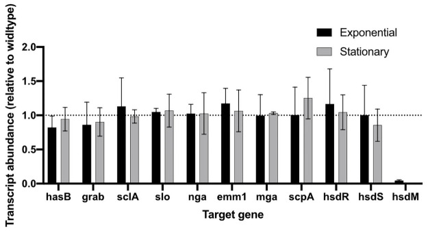
Transcriptional analysis of selected virulence factors in mutant strain 854ΔhsdM.
Data represent mean±SD transcript abundance as determined by qRT-PCR, expressed as a ratio of 854⊗hsdM to wild type strain 854. RNA was isolated from exponential (black) or stationary (gray) phase cultures.
Discussion
In this study we report that the deletion of hsdM in the M1 GAS strain 854 results in increased transformation efficiency due to inactivation of the Hsd restriction modification system. We also observed that the process of transformation of a wild type strain selects for spontaneous mutants in hsdRSM, and that mutations in this locus appear to arise frequently and in a variety of ways. A global analysis of transcript levels in the hsdM deletion strain revealed no significant changes in expression of other genes compared to wild type. A previous study in an M28 strain found that deletion of the entire hsdRSM locus was associated with reduced expression of mga and Mga-regulated genes [2]. That we did not detect changes in transcription of these or other genes may reflect strain- or serotype-specific differences or effects of deleting hsdM alone in contrast to the entire hsd locus.
Various approaches and modifications to restriction-modification systems have been utilized to increase transformation efficiencies in a variety of bacterial species. Inactivation of the Sau1 restriction modification system in Staphylococcus aureus strain RN4220 allows this strain to accept plasmid DNA from E. coli [6]. Plasmid DNA isolated from RN4220 can be further transformed among other S. aureus strains. Modification of a plasmid to eliminate the recognition sequences of restriction modification systems also has resulted in increased ability to transform S. aureus strains without having to use the RN4220 intermediate [7]. Deletion of the gene encoding the restriction endonuclease of a restriction modification system in Caldicellulosiruptor resulted in the ability to take up unmodified plasmid DNA from E. coli and transformation of plasmids isolated from different Caldicellulosiruptor species [8].
In light of the high frequency and variety of naturally occurring mutations we observed in the hsdRSM locus in strains that have undergone transformation in a laboratory setting, we believe it may be advantageous to use a defined ΔhsdM mutant as the parental GAS strain for further genetic manipulations. Use of a defined ΔhsdM mutant such as the one we describe here increases the likelihood of successful transformation, reduces the chance of selecting for spontaneous, uncharacterized hsdRSM mutants, and, in strain 854, appears not to result in significant changes in expression of other genes.
Methods
Bacterial strains and growth conditions
The GAS strain used for molecular manipulation was 854, an M1 strain isolated from a patient with a retroperitoneal abscess [9]. Mutants of strain 854 used in this study are listed in Table 3. GAS was cultured on trypticase soy agar supplemented with 5% defibrinated sheep blood (Remel) or Todd-Hewitt (TH) agar or in TH yeast (THY) broth at 37°C in the presence of 5% CO2. E. coli strain NEB5α (New England Biolabs) was used for cloning. E. coli was cultured in LB broth (Sigma). Growth media were supplemented with spectinomycin at 100 μg/ml when appropriate.
| S. pyogenes strain | Description | Reference |
|---|---|---|
| 854 | Wild type M1 | [9] |
| 854ΔhsdM | 1,005 bp deletion in the coding sequence of hsdM | this study |
| 854ΔcsrS | deletion of csrS | [10] |
| 854CsrS D148N E151Q D152N | Three point mutations in the predicted extracellular domain of CsrS | [11] |
| 854ΔcsrR | Deletion of csrR | [11] |
| 854SLO Y255A | Y255A mutation in SLO | [12] |
| 854Δslo | Deletion of slo | [10] |
| 854speB::Ω | Insertional inactivation of speB | [13] |
| 854CsrS H280A | H280A mutation in CsrS | [11] |
| 854CsrS T284A | T284A mutation in CsrS | [14] |
| 854Δnga | Deletion of nga | [12] |
| 854nga G330D | G330D mutation in NADase | [12] |
Construction of allelic exchange vector and GAS mutagenesis
Oligonucleotide primers are listed in S1 Table. To generate the hsdM deletion mutant, an overlap PCR using Phusion DNA polymerase was performed of the region upstream of hsdM with primers MAB93 and MAB108, and the region downstream of hsdM with primers MAB96 and MAB109. The resulting PCR products were used as template for a second PCR reaction with primers MAB96 and MAB93. The final product was ligated into pJRS233 [15]. The resulting plasmid was transformed into GAS strain 854 by electroporation and subjected to allelic replacement as previously described [16]. Genotype of the mutant strain 854ΔhsdM was confirmed by PCR amplification of the hsdM locus, and phenotype was verified by qRT-PCR for loss of hsdM expression.
RNA isolation and qRT-PCR
GAS were grown in THY broth, and cells were harvested at either mid-exponential (A600 0.5), or early stationary (A600 1) growth phase. Total RNA extraction from bacterial cells was performed using an RNeasy mini kit (Qiagen) according to the manufacturer’s instructions. RNA concentration and purity were determined using a NanoDrop spectrophotometer ND-1000 (Thermo Fisher Scientific). cDNA was generated using the High Capacity RNA-to-cDNA Kit (Applied Biosystems) according to the manufacturer’s instructions.
Quantitative RT-PCR was performed on a QuantStudio 3 System (Applied Biosystems) using the PowerUp SYBER Green Master Mix (Applied Biosystems). Primers are listed in S1 Table. Triplicate assays were performed for each gene tested with 1 ng total template. Expression levels of target genes were normalized to recA (M5005_Spy_1799), and then further normalized to the wild type strain expression levels to obtain a final ΔΔCt value. These values were used to calculate relative expression changes for the mutant strain compared to the parental wild type strain. Data were reported as mean log2 fold-change.
Transformations and quantification of transformation efficiency
Plasmid pDL278 was isolated from E. coli strain NEB5α (New England Biolabs) using a QIAfilter Plasmid Midi Prep (Qiagen). Wild type strain 854 or 854ΔhsdM was subjected to electroporation with 1 μg of plasmid DNA. For preparation of electrocompetent cells, GAS were grown in 20 ml THY broth to A600 of 0.2. Bacterial cells were collected by centrifugation at 4,000 rpm for 10 minutes at 4°C, and then washed with 1 ml ice cold 0.5 M sucrose five times. Cells were resuspended in a final volume of 50 μl 0.5 M sucrose, plasmid DNA was added, and the samples were subjected to electroporation using a Gene Pulser II (Biorad). The parameters used were 1.70 kV, 25 μF, and 400 ohms. After electroporation, each sample was resuspended in 1 ml THY and transferred to a new tube. After one hour of recovery at 37°C, the reaction was serially diluted and spread on TH agar supplemented with 5% defibrinated sheep blood and 100 μg/ml spectinomycin. Colonies, representing transformants, were counted after overnight incubation at 37°C.
RNA extraction for RNA-seq
GAS were grown in THY broth to mid-exponential phase (A600 0.4) and collected by centrifugation. Cell pellets were resuspended in 0.5 mL Trizol reagent (ThermoFisher Scientific) and were transferred to 2 mL FastPrep tubes (MP Biomedicals) containing 0.1 mm Zirconia/Silica beads (BioSpec Products) and shaken for 90 seconds at 10 m/sec speed using the FastPrep-24 5G (MP Biomedicals). After addition of 200 μl chloroform, each sample tube was mixed thoroughly by inversion, incubated for 3 minutes at room temperature, and subjected to centrifugation for 15 minutes at 4°C. The aqueous phase was mixed with an equal volume of 100% ethanol, transferred to a Direct-zol spin plate (Zymo Research), and RNA was extracted according to the Direct-zol protocol (Zymo Research).
Generation of RNA-seq data
Illumina cDNA libraries were generated using a modified version of the RNAtag-seq protocol [17] Briefly, 0.5 to 1 μg of total RNA was fragmented, depleted of genomic DNA, dephosphorylated, and ligated to DNA adapters carrying 5’-AN8-3’ barcodes of known sequence with a 5’ phosphate and a 3’ blocking group. Barcoded RNAs were pooled and depleted of rRNA using the RiboZero rRNA depletion kit (Epicentre). Pools of barcoded RNAs were converted to Illumina cDNA libraries in 2 main steps: (i) reverse transcription of the RNA using a primer designed to the constant region of the barcoded adapter with addition of an adapter to the 3’ end of the cDNA by template switching using SMARTScribe (Clontech) as described [18] (ii) PCR amplification using primers whose 5’ ends target the constant regions of the 3’ or 5’ adapters and whose 3’ ends contain the full Illumina P5 or P7 sequences. cDNA libraries were sequenced on the Illumina [Nextseq 2500] platform to generate paired-end reads.
RNA-seq data analysis
Paired-end sequencing reads were mapped to the S. pyogenes MGAS5005 genome (NCBI RefSeq NC_007297) using bowtie2 version 2.3.1 [19]. Reads that mapped to annotated genes were counted using HTSeq version 0.10.0 and analysis of differential gene expression was conducted using DESeq2 version 1.20.0. Reported genes had a two-fold greater or lesser abundance than wild type, all with an adjusted P-value of 0.05 or less.
Modeling of HsdM from GAS
Sequence of HsdM from MGAS5005 (accession number WP_010922657.1) was submitted to I-TASSER to be compared with the cryo-EM structure of HsdR2M2S1-OcrIS from EcoR124I (PBD 7BST) [20,21]. Molecular graphics and analyses were performed with UCSF Chimera, developed by the Resource for Biocomputing, Visualization, and Informatics at the University of California, San Francisco [22].
Acknowledgements
We thank Jorge Velarde for assistance in modeling the GAS HsdM structure and generating the structural figure. RNA-seq libraries were constructed and sequenced at the Broad Institute of MIT and Harvard by the Microbial ‘Omics Core and Genomics Platform, respectively.
References
1
2
3
4
5
6
7
8
9
10
11
12
13
14
15
16
17
18
19
20
21
22